
Amniocentesis
Amniocentesis is a medical procedure performed during pregnancy to obtain a small sample of amniotic fluid. This fluid surrounds the fetus in the womb and contains cells and other substances that can provide valuable information about the health of the fetus.
During the procedure, which is usually performed between weeks 15 and 20 of pregnancy, a thin needle is inserted through the abdomen into the amniotic sac under ultrasound guidance. A small amount of amniotic fluid, typically around 20 milliliters, is then withdrawn and sent to a laboratory for analysis.
Amniocentesis can be used for various purposes, including:
- Genetic testing: It can detect chromosomal abnormalities such as Down syndrome (trisomy 21), Edwards syndrome (trisomy 18), and Patau syndrome (trisomy 13). It can also diagnose genetic disorders such as cystic fibrosis and sickle cell disease.
- Fetal lung maturity: In some cases, particularly when a preterm delivery is being considered, amniocentesis can determine whether the baby’s lungs are mature enough for birth.
- Infections: It can detect infections in the fetus, such as toxoplasmosis or cytomegalovirus, which can affect the baby’s health.
- Rhesus (Rh) factor: It can determine the baby’s blood type and whether the baby is at risk for developing Rh disease, a condition in which a mother’s antibodies attack the baby’s red blood cells.
When is amniocentesis performed?
Amniocentesis is typically performed between weeks 15 and 20 of pregnancy, usually around the 16th week, but it can be done later in some cases. This timing is chosen because it provides sufficient amniotic fluid for sampling while minimizing risks to the pregnancy. However, it can be performed earlier in cases of medical necessity.

How should I prepare for an amniocentesis?
Preparing for an amniocentesis involves both physical and mental preparation. Here are some steps to follow:
- Consultation with Your Doctor: Before the procedure, have a thorough discussion with your doctor about why you need the amniocentesis, what it involves, and what the risks and benefits are. This will help alleviate any concerns and ensure you’re fully informed.
- Timing: Schedule the amniocentesis at the appropriate time in your pregnancy. Typically, this is done between weeks 15 and 20.
- Hydration: Drink plenty of fluids before the procedure. This can help increase the volume of amniotic fluid and make it easier to obtain a sample.
- Rest: Ensure you’re well-rested before the procedure. Fatigue can make any medical procedure more challenging.
- Clothing: Wear comfortable clothing that allows easy access to your abdomen. You may be more comfortable in loose-fitting clothing.
- Avoid Aspirin and Blood Thinners: If you’re taking any medications like aspirin or other blood thinners, consult with your doctor about whether you need to stop taking them temporarily before the procedure.
- Bring Support: It’s a good idea to bring someone with you for support, both during and after the procedure. This could be a partner, friend, or family member.
- Emotional Support: Acknowledge any fears or anxieties you may have and discuss them with your healthcare provider. Sometimes, simply talking about concerns can help alleviate them.
- Know the Risks: Understand the potential risks of the procedure, such as infection, leakage of amniotic fluid, and miscarriage (though this is rare, occurring in about 1 in 300 to 1 in 500 procedures).
- Plan for Rest Afterwards: Arrange for some rest time after the procedure. While most women feel little discomfort, some may experience cramping or mild discomfort, so it’s wise to plan for some downtime.
- Ask Questions: If you have any doubts or questions, don’t hesitate to ask your healthcare provider. It’s essential to have a clear understanding of what to expect.
What are the risks of amniocentesis?
While it’s generally considered safe, there are some risks associated with the procedure:
- Miscarriage: The most significant risk associated with amniocentesis is miscarriage, which occurs in about 0.3-0.6% of cases. This risk is slightly higher if the procedure is done earlier in pregnancy.
- Infection: There’s a small risk of infection following amniocentesis, which can lead to complications for both the mother and the fetus. This risk is minimized by using sterile techniques during the procedure.
- Leakage of amniotic fluid: In rare cases, the needle used to collect the fluid can cause fluid leakage from the amniotic sac. This can lead to complications such as premature labor or infection.
- Rh sensitization: If the mother is Rh-negative and the fetus is Rh-positive, there’s a risk of Rh sensitization, where the mother’s immune system may produce antibodies against the fetus’s blood cells. This can cause complications in future pregnancies.
- Needle injury: There’s a small risk of injury to the fetus if the needle accidentally comes into contact with the fetus during the procedure. However, ultrasound guidance is usually used to minimize this risk.
- Discomfort: Some women may experience discomfort or pain during the procedure, usually mild and temporary.
How do doctors perform amniocentesis?
- Preparation: The doctor typically starts by performing an ultrasound to locate the position of the fetus and the placenta. This helps in determining a safe insertion site for the needle.
- Positioning: The pregnant woman is usually asked to lie down on her back, although in some cases, a different position may be preferred depending on factors such as the position of the uterus.
- Sterilization: The area of the abdomen where the needle will be inserted is cleaned and sterilized to reduce the risk of infection.
- Numbing the area: A local anesthetic may be injected into the skin to numb the area where the needle will be inserted. This helps to minimize discomfort during the procedure.
- Inserting the needle: Using ultrasound guidance to visualize the fetus and the placenta, the doctor carefully inserts a thin, hollow needle through the abdominal wall and into the amniotic sac.
- Collecting the fluid: Once the needle is properly positioned within the amniotic sac, a small amount of amniotic fluid (usually around 20 milliliters) is drawn into a syringe attached to the needle.
- Removing the needle: After the fluid sample is collected, the needle is carefully removed.
- Monitoring: The woman is usually monitored for a short period after the procedure to ensure there are no immediate complications, such as bleeding or contractions.
- Follow-up: After the procedure, the amniotic fluid sample is sent to a laboratory for analysis. Results are typically available within a few days to a couple of weeks, depending on the tests being performed.
How painful is amniocentesis?
Amniocentesis is generally associated with some discomfort, but the level of pain varies from person to person. During the procedure, a thin needle is inserted through the abdomen into the uterus to withdraw a small amount of amniotic fluid for testing.
Most people describe the sensation as a sharp pinch or cramp similar to menstrual cramps, but it usually lasts only for a short time, typically less than a minute. Some women report feeling pressure or mild discomfort as the needle is inserted, but it’s usually manageable.
What do the results of an amniocentesis test mean?
- Chromosomal Abnormalities: One of the primary reasons for performing amniocentesis is to detect chromosomal abnormalities such as Down syndrome (Trisomy 21), Edwards syndrome (Trisomy 18), or Patau syndrome (Trisomy 13). The results will indicate whether the fetus has the usual number of chromosomes (46) or if there is an extra chromosome present (trisomy) or a missing chromosome (monosomy).
- Genetic Disorders: Amniocentesis can also detect specific genetic disorders caused by mutations in genes. These disorders include cystic fibrosis, sickle cell disease, and Tay-Sachs disease, among others.
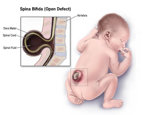
- Neural Tube Defects: The test can identify neural tube defects such as spina bifida or anencephaly. These are structural defects in the spinal cord or brain.
- Lung Maturity: In some cases, amniocentesis is performed to assess fetal lung maturity, particularly if there’s a risk of preterm birth. Lung maturity is crucial for a baby’s ability to breathe outside the womb.
- Infection: Amniocentesis can also detect infections, such as toxoplasmosis, cytomegalovirus (CMV), or Zika virus, which can affect the fetus.
[…] insufficiency, is a disorder in which the adrenal glands are unable to generate adequate DHEAS. Fatigue, weakness in the muscles, loss of weight, low blood pressure, and skin discoloration are some of […]
[…] heel for these tests. The main objectives are identifying viral infections and metabolic, genetic, and endocrine […]
[…] process of amniocentesis is used to extract amniotic fluid. A tiny amount of fluid is removed by inserting a needle, guided […]
[…] the presence of an extra copy of chromosome 21. Instead of the typical two copies, individuals with trisomy 21 have three copies of this chromosome. This extra genetic material affects the development of the […]
[…] Villus Sampling (CVS) and amniocentesis are prenatal tests to detect genetic abnormalities in a developing […]
[…] Antibody Test, This test is done in those pregnant women whose spouses have RH type (RH type) which is not compatible with each other i.e. one has […]